Modern dentistry is unthinkable without X-ray imaging.
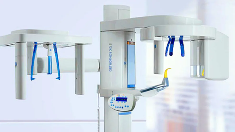
Today, the “Nino Beridze Orthodontic Center” presents something new: a unit from the German company Sirona for orthopantomography and cephalometric imaging.
For more than 25 years, Sirona’s dental systems have held a leading position in the innovative development of dental technology worldwide. Sirona keeps up with dental market trends and consistently offers top-level dental equipment.
With it, the problem in each specific case can be precisely identified.
Diagnosis is the foundation of predictable treatment! To get complete dental treatment, the patient’s whole mouth is examined.
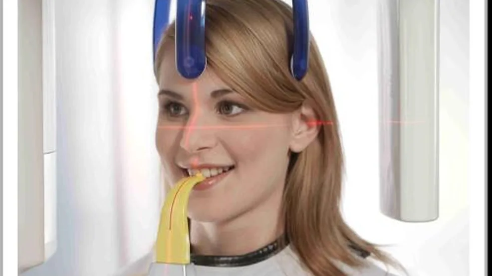
As the saying goes: “Whoever diagnoses well, treats well.” At our clinic, diagnostics are carried out with an X-ray machine from the German company Sirona. With this device, the radiation dose is 80% lower than with traditional machines.
The Orthopos XG 5 has 12 digital panoramic imaging programs.
The device is equipped with a special program for children.
Complete diagnostics are essential before orthodontic treatment. Diagnostics may include various examination methods used to determine the bite, and additional tests may also be needed.
Orthopantomogram –
A single-exposure image of the entire dental and jaw system as one functional complex.
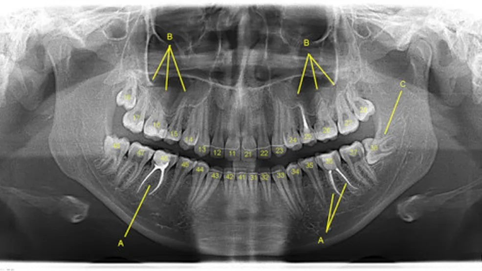
The image covers the alveolar ridges in full, and the image itself is laid out on a single plane. With minimal radiation exposure for the patient, an orthopantomogram gives a high-quality, complete picture on a large scale. The image shows:
- the main cavities;
- bone tissue;
- pathology and its visible boundaries;
- the position of the roots.
Orthopantomograms are used by various specialists: orthodontists, implantologists, general dentists and prosthodontists. In some cases, surgeons simply can’t work without one.
Cephalometry – this examination is used to diagnose various bite anomalies, confirm the accuracy of a diagnosis and predict the outcome of orthodontic treatment. Images of the facial and cranial skull, as well as soft tissue contours, make it possible to determine how they relate to each other and to analyze the structure of the facial skeleton.
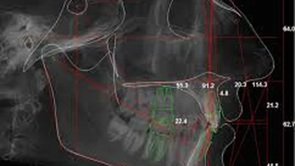
Teleradiography (TRG) – (Greek tele – far, + radiography) – radiography performed from a long distance to obtain an X-ray image with the most accurate dimensions of the object being examined. This method is widely used in orthodontics. Its data are essential for an orthodontic diagnosis and correct treatment. TRG is always performed before orthodontic treatment, or during treatment as indicated, and at the end of treatment. A teleradiograph is a lateral or frontal image of the skull and is used for cephalometry – very precise calculations of the relationships between different areas of the facial skeleton. TRG is used to determine the correct position of the upper and lower jaws in relation to the skull and the inclination of the teeth, as well as to assess the soft tissues of the face. The image obtained with teleradiography shows every tissue of the area in question in the finest detail.
The examinations listed above can be carried out on referral from a dentist, once the method and its importance for starting treatment have been determined.
Based on the examinations, doctors draw up a plan for the treatment and procedures to be carried out. For X-ray examinations, you need to see a specialist who will prescribe the examination method you need along with a treatment plan.